")
Angel of the West (This sculpture represents an IgG molecule)
Immunoglobulins are glycoproteins that function as antibodies. In fact, the terms antibodies and immunoglobulins are usually used indistinctly: immunoglobulins highlight structure and antibody highlights function. Immunoglobulins can be found attached to the B-cell membranes, in secretions or circulating in blood.
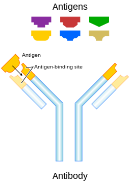
Immunoglobulins are produced as a response to the detection of foreign molecules in our body. These foreign substances that trigger the production of antibodies are called antigens.
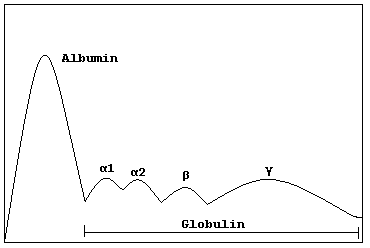
Circulating immunoglobulins are included in the plasma protein fraction of the gamma globulins.
There are different types of immunoglobulins: IgG, IgM, IgA, IgD and IgE. All of them have in common that their basic unit is formed by two pairs of peptide chains: a pair of Light chains or L chains (approximately 220 amino acids each) and a pair of Heavy chains or H chains (around 440 amino acids each).
These four chains in the basic structure are linked through disulfide bridges between cystein residues in the backbone of the peptide chains. Each Light chain is linked to one Heavy chain and each Heavy chain is associated to a Light chain and to the other Heavy chain.
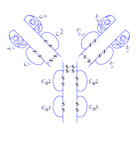
The following graphic shows the Heavy chains in blue, the Light chains in green and the disulfide linkages between the chains in red (there are additional intrachain disulfide bridges that are not shown in this graphic)
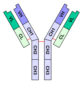
Observe also in the graphic that in the L chains can be distinguished two regions or domains: VL and CL, while in the H chains, 4 regions or domains can be found: VH, CH1, CH2 and CH3. Each of these regions is composed by 70 to 110 amino acids.
The V regions are Variable regions: the amino acid sequence in these regions (the NH2- terminal regions of L and H chains) is highly variable, and within them, in the L and in the C chains, there are hyper variable regions (CDRs of Complementarity-determining regions) that form the specific antigen binding site complementary to the specific antigen.
This video shows the structure of a typical immunoglobulin IgG:
As you have seen, there are two binding sites for antigens in each (LH)2 unit. When a (LH)2 unit is hydrolyzed with papain, three fragments are released: two Fab and one Fc fragment.
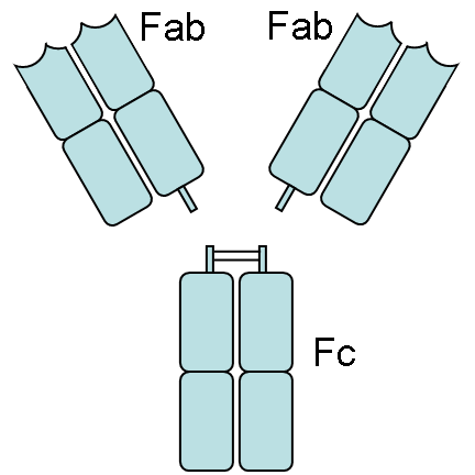
The Fab fragments contain the structure that is able to bind to the antigen (Fab = Fragment antigen-binding), while the Fc fragment (c means crystallizable) is not able to bind to the antigen, but contain a complement binding site, that is exposed when the interaction between the Fab fragment and the antigen occurs. This binding occurs through non covalent interactions (Van der Waals forces, Hydrogen bonds, hydrophobic interactions) and triggers conformational changes similar to those observed in the enzyme-substrate inducing fit mechanism. This allosteric effect exposes sites in the constant regions of the heavy chains, related to the binding and activation of complement proteins.
This complement system is formed by eleven different proteins that are sequentially activated for associating to the cell membrane to cause lysis and death of the invading bacterial cell.
Another important role of the complement system is to generate proteins called opsonins, which stimulate phagocytosis by neutrophils and macrophages.
(More detailed information about the complement system can be found here)
In addition to the activation of the complement system, the constant regions of the Heavy chains define the ability of the (LH)2 basic structure to associate to others (LH)2 units and determine the kind of immunoglobulin.
There are four kind of Heavy chains: g (Gamma), a (Alpha), d (Delta), e (Epsilon) and m (Mu).
Gamma chains are similar in their constant regions that are different to the other kind of heavy chain constant regions. The same is valid for each of the different kinds of Heavy chains.
Immunoglobulins that contain Gamma chains are called IgG. IgG molecules are formed by one (LH)2 unit. These are the most abundant immunoglobulins in sera (600-1800 mg/dL). They promote phagocytosis in plasma and activate the complement system. IgG are the only kind of antibodies that can cross the placenta.
Observe in the following diagram of an IgG molecule, the two Heavy chains (in red and blue) and the two Light chains (in green and yellow).

In this diagram can be observed the variable and constant regions of the IgG and the interchain and intrachain disulfide linkages in the structure.
Immunoglobulins containing alpha chains are called IgA. IgA is found mainly in mucosal secretions, tears, colostrum and milk. These are the initial defense in mucosas against pathogen agents. They appear usually as dimmers of (LH)2 units
IgM contain mu heavy chains. IgM antibodies are expressed in the surface of B-cells and are found primarily in plasma. They are the first antibodies produced in significant quantities against an antigen. They promote phagocytosis and activate the complement system. They appear usually as pentamers of (LH)2 units

Ig E contains Heavy chains type epsilon. IgE, a (LH)2 monomer, plays an important role in allergic reactions and increase in worm infestations.
The role of IgD (immunoglobulins with delta heavy chains) is not very well known. This kind if IgG is found in the surface of the B-cells that have not been exposed to antigens. IgD structure correspond also to a (LH)2 monomer,
There are also different classes of L chains: the Lambda (l) and Kappa (k) class. Each immunoglobulin molecule has either lambda or kappa chains, but not both.
Lambda chain are similar in their constant regions, Kappa chains are similar between them in those regions.
In summary, immunoglobulins are proteins that function as antibodies. The basic structure of immunoglobulins is a unit formed by two light chains and two heavy chains. These units contain variable domains and constant domains. The variable domains of the L and H chains are responsible of the binding to the antigens, while the constant regions of the H chains are responsible for the activation of the complement system and the ability of some of these (LH)2 units to form polymers.

i want to know wat is fab.
fab is the fragment for antiggen binding, Antibody prform two functions, one is to bind specifically to molecules from the pathogens, and second is to recruit other cells and molecules to destroy the pathogen..
in tis page i didnt understood wat is fab
i am very interested on this site and thanks for website provider.
its nice presentation,really superb,please explain bencejones proteins,and what happend if lambda chain ,kappa hain exess will occur
awesome. Cleared the functions of the different immunoglobulins for me! Thanks!
This is excellent,sufficient,useful,imp,and easy to understand 4 me.
This is excellent,sufficient,useful,imp, easy to understand,and nice presentation,really
superb
its nice presentation,really
superb,thanks for website
provider.
Fantastic clear lucid elegant systematic logical explanation of immunoglobulins. Many many thanks. If the texbooks were so very lucid, Imwould notmhave to search for help!
i want to know,are there different different structures for every immunoglobulin like IgG,IgM,IgA,IgD and IgE……?yhen please show the structures.
want to know more about antibodies nd their functions in detail nd latest reseach on these??????? pls tell more
nice,but is there a link where I can find the genetic rearrangement of immunoglobulins in a simple and understandable way like this one.
can u pls tell me about the scope of biochem in coming days??
nice
Thanks for such an educating write up. Please, tell me how are antibodies synthesized? include the genes coding for the proteins and the mechanism.